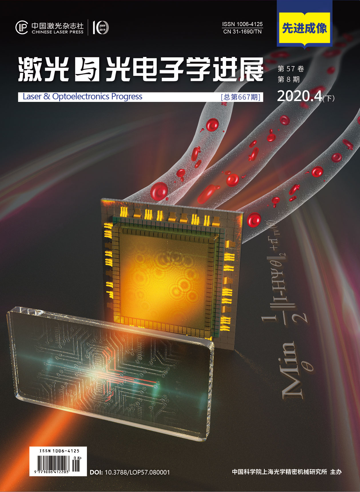
激光与光电子学进展, 2020, 57 (8): 080002, 网络出版: 2020-04-03
高光谱在体组织成像方法的研究进展  下载: 1736次
Hyperspectral Imaging of in vivo Tissues: A Review
成像系统 高光谱成像 生物医学光子学 组织光学成像 imaging systems hyperspectral imaging biomedical photonics tissue optical imaging
摘要
高光谱成像方法可同时获取在体组织的二维图像与光谱信息,具有空间与光谱分辨率高、成像范围大、无损快速等优点,为在体诊断提供了丰富的信息。近年来,研究人员在成像方法、仪器与应用方面进行了大量研究,取得了长足进展。本文综述了高光谱在体组织成像方法及应用研究的主要进展,探讨高光谱成像分光方法、系统组成与特点。从光谱重构方法、组织光学参数测量、基于深度学习的图像处理方法等方面,介绍在体组织成像与成分检测方法的研究进展。同时,对高光谱成像在临床医学,如皮肤创伤与愈合过程检测,糖尿病足与视网膜疾病诊断,手术中检测及微循环功能评估等方面的应用进展进行了总结。
Abstract
Hyperspectral imaging method can obtain two-dimensional images and spectral information of in vivo tissues, which has the advantages of high spatial and spectral resolution, large imaging range, non-invasive nature, and fast speed, providing abundant information for in vivo tissue diagnosis. In recent years, researchers have made great progress in imaging methods, instruments, and applications. In this study, the main advances of hyperspectral imaging methods and applications are reviewed, and the spectral imaging methods, system composition, and characteristics are discussed. This study introduces the research progress in in vivo tissue imaging methods and application in terms of spectral reconstruction, tissue optical parameter measurement, and image processing based on deep learning. Simultaneously, the application progress of hyperspectral imaging in clinical medicine, such as skin trauma and healing process detection, diagnosis of diabetic foot and retinal diseases, intraoperative detection, and microcirculation function evaluation, is also summarized.
马雪洁, 刘蓉, 李晨曦, 陈文亮, 徐可欣. 高光谱在体组织成像方法的研究进展[J]. 激光与光电子学进展, 2020, 57(8): 080002. Xuejie Ma, Rong Liu, Chenxi Li, Wenliang Chen, Kexin Xu. Hyperspectral Imaging of in vivo Tissues: A Review[J]. Laser & Optoelectronics Progress, 2020, 57(8): 080002.


