中国激光, 2020, 47 (2): 0207029, 网络出版: 2020-02-21
β-淀粉样蛋白斑块的无标记成像及光动力降解  下载: 1252次
Label-Free Imaging of β-Amyloid Plaques and Photodynamic Degradation
图 & 表
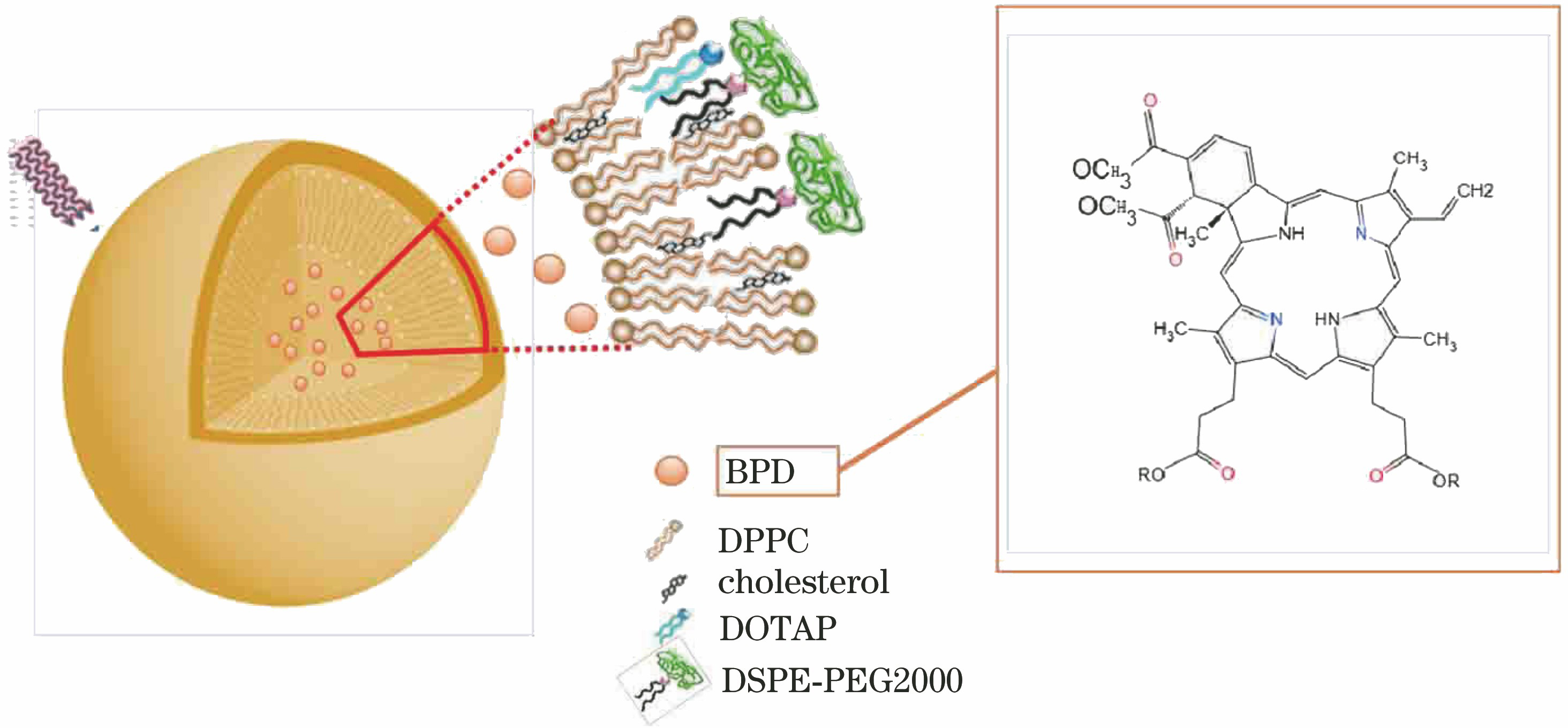
图 2. Aβ溶液的不同聚集状态。(a) Aβ纤维;(b) Aβ球晶;(c) Aβ斑块
Fig. 2. Different aggregation states of Aβ solution. (a) Aβ fibers; (b) Aβ spherulites; (c) Aβ plaques

图 3. 不同质量浓度下BPD溶液的紫外吸收及荧光发射谱。(a) BPD紫外吸收谱;(b) BPD吸光度随质量浓度变化的标准曲线;(c) BPD荧光发射谱;(d) BPD荧光发射强度随质量浓度变化的标准曲线
Fig. 3. Absorption and fluorescence emission spectra of BPD solution with different mass concentrations. (a) BPD ultraviolet absorption spectra; (b) standard curve of BPD absorbance varying with mass concentration; (c) BPD fluorescence emission spectra; (d) standard curve of BPD fluorescence intensity varying with mass concentration

图 4. BPD及其脂质体对Aβ聚集体的降解机理
Fig. 4. Degradation mechanism of Aβ aggregates by BPD and its liposomes

图 5. Aβ斑块在仅光照情况下的降解结果。 (a)(e)明场图;(b)(f)共聚焦成像图;(c)(g)二次谐波成像图;(d)(h)双光子荧光成像图
Fig. 5. Degradation results of Aβ plaques with only irradiation. (a)(e) Bright field images; (b)(f) confocal images; (c)(g) second harmonic generation images; (d)(h) two photon excited fluorescence images

图 6. BPD浓度为1 μmol/L情况下光照Aβ斑块的降解结果。(a)(e)明场图;(b)(f)共聚焦成像图;(c)(g)二次谐波成像图;(d)(h)双光子荧光成像图
Fig. 6. Degradation results of Aβ plaques after irradiation when the concentration of BPD is 1 μmol/L. (a)(e) Bright field images; (b)(f) confocal images; (c)(g) second harmonic generation images; (d)(h) two photon excited fluorescence images

图 7. BPD浓度为10 μmol/L情况下光照Aβ斑块的降解结果。(a)(e)(i)(m)明场图;(b)(f)(j)(n)共聚焦成像图;(c)(g)(k)(o)二次谐波成像图;(d)(h)(l)(p)双光子荧光成像图
Fig. 7. Degradation results of Aβ plaques after irradiation when the concentration of BPD is 10 μmol/L. (a)(e)(i)(m) Bright field images; (b)(f)(j)(n) confocal images; (c)(g)(k)(o) second harmonic generation images; (d)(h)(l)(p) two photon excited fluorescence images

图 8. Aβ斑块荧光强度随光照时间的变化图
Fig. 8. Relationship between fluorescence intensity of Aβ plaques and irradiation time

图 9. BPD浓度为100 μmol/L情况下光照Aβ斑块的降解结果。(a)(e)明场图;(b)(f)共聚焦成像图;(c)(g)二次谐波成像图;(d)(h)双光子荧光成像图
Fig. 9. Degradation results of Aβ plaques after irradiation when the concentration of BPD is 100 μmol/L. (a)(e) Bright field images; (b)(f) confocal images; (c)(g) second harmonic generation images; (d)(h) two photon excited fluorescence images

图 10. BPD对Aβ斑块的降解效率随浓度的变化关系
Fig. 10. Relationship between degradation efficiency of Aβplaques and concentration of BPD

图 11. 不同剂量光照后Aβ含量变化的ELISA检测结果
Fig. 11. ELISA detection results of Aβ content after irradiation with different doses

图 12. 加入BPD脂质体情况下光照Aβ斑块的降解结果。 (a)(e)明场图;(b)(f)共聚焦成像图;(c)(g)二次谐波成像图;(d)(h)双光子荧光成像图
Fig. 12. Degradation results of Aβ plaques after irradiation with BPD liposome. (a)(e) Bright field images; (b)(f) confocal images; (c)(g) second harmonic generation images; (d)(h) two photon excited fluorescence images

黄燕霞, 许皓, 栾萍, OhulchanskyyTymishY, 屈军乐. β-淀粉样蛋白斑块的无标记成像及光动力降解[J]. 中国激光, 2020, 47(2): 0207029. Huang Yanxia, Xu Hao, Luan Ping, Ohulchanskyy Tymish Y, Qu Junle. Label-Free Imaging of β-Amyloid Plaques and Photodynamic Degradation[J]. Chinese Journal of Lasers, 2020, 47(2): 0207029.


 PDF全文
PDF全文

