光学相干层析成像图像中角膜厚度的自动测量方法  下载: 1254次
Automatic Measurement Method for Corneal Thickness of Optical Coherence Tomography Images
1 上海大学机电工程与自动化学院, 上海 200444
2 中国科学院上海光学精密机械研究所信息光学与光电技术实验室, 上海 201800
图 & 表
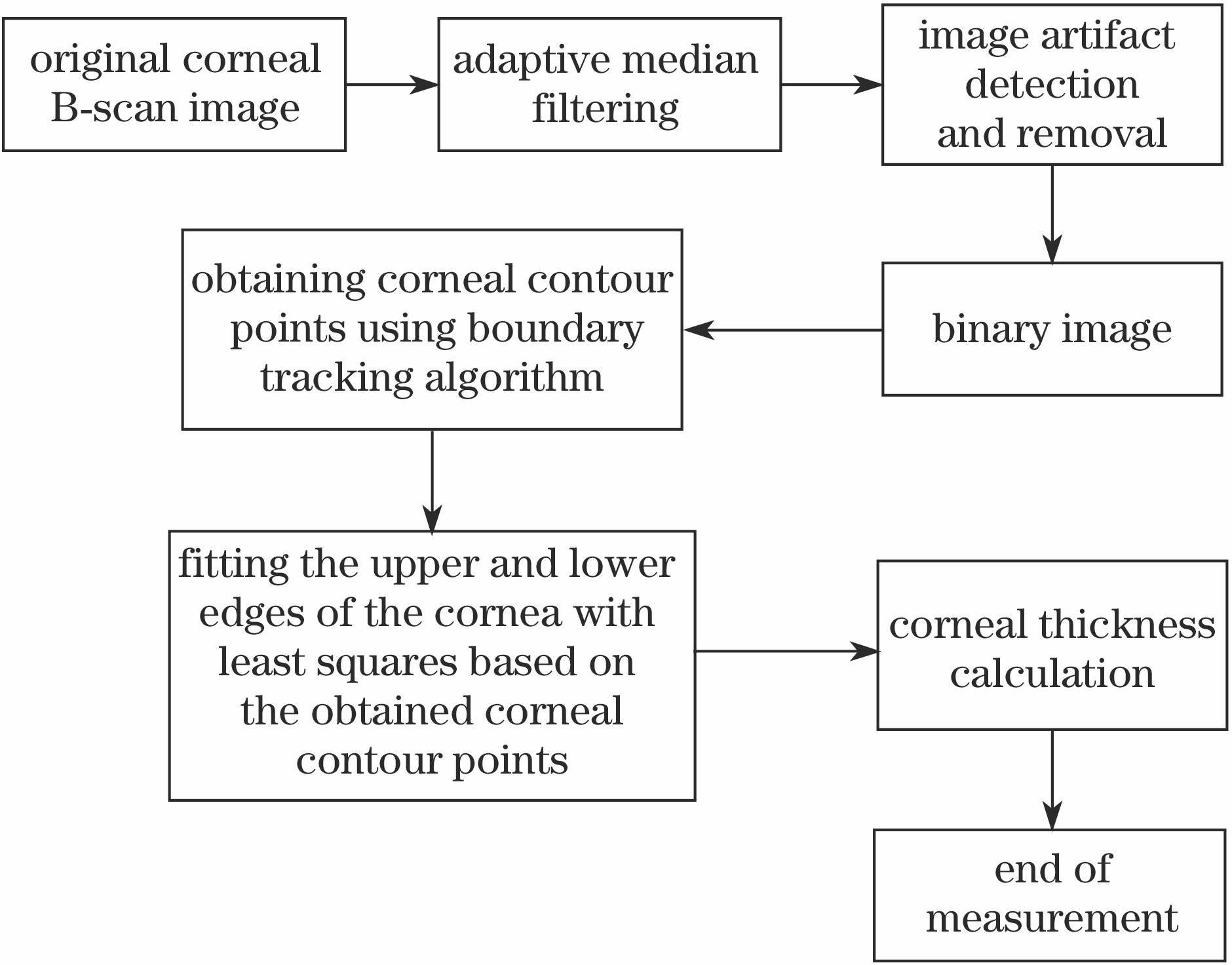
图 1. 所提角膜厚度测量方法流程图
Fig. 1. Flow chart of proposed measurement method of corneal thickness
下载图片 查看原文
图 2. 原始角膜B扫描图像中存在的噪声和伪影
Fig. 2. Noise and artifacts in original cornea B scan image
下载图片 查看原文
图 3. A-扫描平均强度曲线
Fig. 3. A-scan averaged intensity curve
下载图片 查看原文
图 4. 相邻行平均强度曲线
Fig. 4. Average intensity curves of adjacent rows
下载图片 查看原文
图 5. 角膜B扫描OCT图像经过预处理后的效果。(a)原始角膜B扫描图像;(b)自适应中值滤波后的角膜B扫描图像;(c)去除水平伪影后的角膜B扫描图像;(d)抑制中央伪影的角膜B扫描图像
Fig. 5. Effect of corneal B-scan OCT images after pre-treatment. (a) Original corneal B-scan image; (b) corneal B-scan image with adaptive median-filtering; (c) corneal B-scan image with horizontal artifact removal; (d) corneal B-scan image with suppressed central artifact
下载图片 查看原文
图 6. 边界条件。(a)外边界;(b)孔边界
Fig. 6. Boundary conditions. (a) Outer boundary condition; (b) hole boundary condition
下载图片 查看原文
图 7. 人眼角膜B扫描OCT图像上下边缘的拟合结果。(a)高质量的原始角膜B扫描图像;(b)高质量的角膜B扫描图像的边缘拟合结果;(c)存在噪声和伪影的原始角膜B扫描图像;(d)存在噪声和伪影的角膜B扫描图像的边缘拟合结果
Fig. 7. Upper and lower edge fitting results of human corneal OCT B-scan image. (a) High quality original corneal B-scan image; (b) high quality edge fitting corneal B-scan image; (c) original corneal B-scan image with noise and artifacts; (d) edge fitting corneal B-scan image with noise and artifacts
下载图片 查看原文
图 8. 不同预处理去噪算法得到的人眼角膜B扫描图像上下边缘的拟合效果。(a)未进行预处理去噪;(b)均值滤波预处理;(c)中值滤波预处理;(d)自适应中值滤波预处理
Fig. 8. Fitting effects of upper and lower edges of human corneal B-scan images obtained by different pre-processing denoising algorithms. (a) No preprocessing denoising. (b) mean filter preprocessing; (c) median filter preprocessing; (d) adaptive median filter preprocessing
下载图片 查看原文
图 9. OCT角膜图像上下边缘的拟合结果。(a)边缘检测和随机抽样一致性方法;(b)所提方法
Fig. 9. Results of upper and lower edge fitting of OCT corneal images. (a) Edge detection and random sampling consistency method; (b) proposed method
下载图片 查看原文
表 1不同质量角膜图像沿Y轴方向的平均厚度、中央厚度和对应的标准方差
Table1. Average thickness, central thickness and corresponding standard deviations along Y-axis of corneal images with different qualities
| Algorithm type | Ta±σ /μm | Tc±σ /μm |
|---|
| High quality corneal B-scan image | 561.6±1.2 | 571.6±2.9 | | Corneal B-scan image with noise and artifacts | 562.1±2.3 | 572.3±3.8 |
|
查看原文
表 2采用不同预处理算法处理后角膜沿Y轴方向的平均厚度、角膜中央厚度和对应的标准方差
Table2. Average thickness, central thickness and corresponding standard deviations along Y-axis after treatments with different pretreatment algorithms
| Preprocessing algorithm | Ta±σ /μm | Tc±σ /μm |
|---|
| No preprocessing denoising | 567.2±5.1 | 575.3±5.4 | | Mean filter preprocessing | 563.1±4.4 | 573.9±4.8 | | Median filter preprocessing | 564.6±3.8 | 573.2±4.5 | | Adaptive median filter preprocessing | 562.1±2.3 | 572.3±3.8 | | Manual measurement | 561.4±1.4 | 571.2±2.4 |
|
查看原文
表 3不同方法下角膜沿Y轴方向的平均厚度偏差、角膜中心厚度偏差和对应的标准方差
Table3. Average thickness deviation, corneal center thickness deviation and corresponding standard deviations along Y-axis of corneal by different methods
| Algorithm type | Proposed algorithm (manual measurement) | Random sampling consisitency (RANSC) algorithm (manual measurement) |
|---|
| Ta±σ /μm | 1.0±0.3 | 2.3±1.8 | | Tc±σ /μm | 1.2±0.6 | 5.2±3.9 |
|
查看原文
高阳, 李中梁, 张建华, 南楠, 王瑄, 王向朝. 光学相干层析成像图像中角膜厚度的自动测量方法[J]. 光学学报, 2019, 39(3): 0311003. Yang Gao, Zhongliang Li, Jianhua Zhang, Nan Nan, Xuan Wang, Xiangzhao Wang. Automatic Measurement Method for Corneal Thickness of Optical Coherence Tomography Images[J]. Acta Optica Sinica, 2019, 39(3): 0311003.


 PDF全文
PDF全文

