
Author Affiliations
Abstract
1 College of Engineering and Applied Sciences, Nanjing University, Nanjing 210093, China
2 China Academy of Electronics and Information Technology, Beijing 100041, China
A metal-lined hollow-core fiber-based Raman probe extension kit is proposed in this Letter for in situ and sensitive ultramicro-analysis. A hollow-core fiber can confine light and fluid samples in its hollow core, with enhanced light–sample interaction. By using a homemade light coupling device with a glass window for liquid isolation, a 3.5-cm-long hollow-core fiber could mount on and connect to a Raman probe, with perfect light coupling efficiency. After full filling the hollow-core fiber chamber with a volume of 13 μL by using a syringe pump, it can act as an extension kit for an ordinary Raman probe and be used as a ultramicro-analysis tool for the sample of microfluidic chips. In order to enhance its sensitivity, a gold film coated fiber tip is inserted into the capillary, which can double the Raman signal received by reflecting pump light and Raman light. Finally, a detection limit of 5% for ethanol solution and an enhancement factor of two compared with direct detection of bulk sample volume are demonstrated. Above all, our device can be utilized as a Raman probe extension kit, which is suitable for rapid, sensitive, and in situ measurements for a few microliter level samples.
060.2370 Fiber optics sensors 300.6450 Spectroscopy, Raman Chinese Optics Letters
2019, 17(11): 110601

Author Affiliations
Abstract
1 Department of Electrical and Systems Engineering, Washington University in St. Louis, St. Louis, Missouri 63130, USA
2 Department of Electrical Engineering, Pennsylvania State University, University Park, Pennsylvania 16802, USA
Conventionally, metallic nanostructures are used for surface-enhanced Raman spectroscopy (SERS), but recently there has been increasing interest in the enhancement of Raman scattering from dielectric substrates due to their improved stability and biocompatibility compared with metallic substrates. Here, we report the observation of enhanced Raman scattering from rhodamine 6G molecules coated on silica microspheres. We excite the whispering gallery modes (WGMs) supported in the microspheres with a tapered fiber coupler for efficient WGM excitation, and the Raman enhancement can be attributed to the WGM mechanism. Strong resonance enhancement in pump laser intensity and modified Raman emission from the Purcell effect in the microsphere resonator are observed from the experiment and compared with theoretical results. A total Raman enhancement factor of 1.4×10⁴ is observed, with contribution mostly from the enhancement in pump laser intensity. Our results show that, with an efficient pumping scheme, dielectric microspheres are a viable alternative to metallic SERS substrates.
Spectroscopy, Raman Surface-enhanced Raman scattering Resonators Photonics Research
2018, 6(5): 05000346
Author Affiliations
Abstract
1 State Key Laboratory of Optoelectronic Materials and Technologies, School of Materials, Sun Yat-sen University, Guangzhou 510275, China
2 Key Laboratory of Materials Physics, Institute of Solid State Physics, Chinese Academy of Sciences, Hefei 230031, China
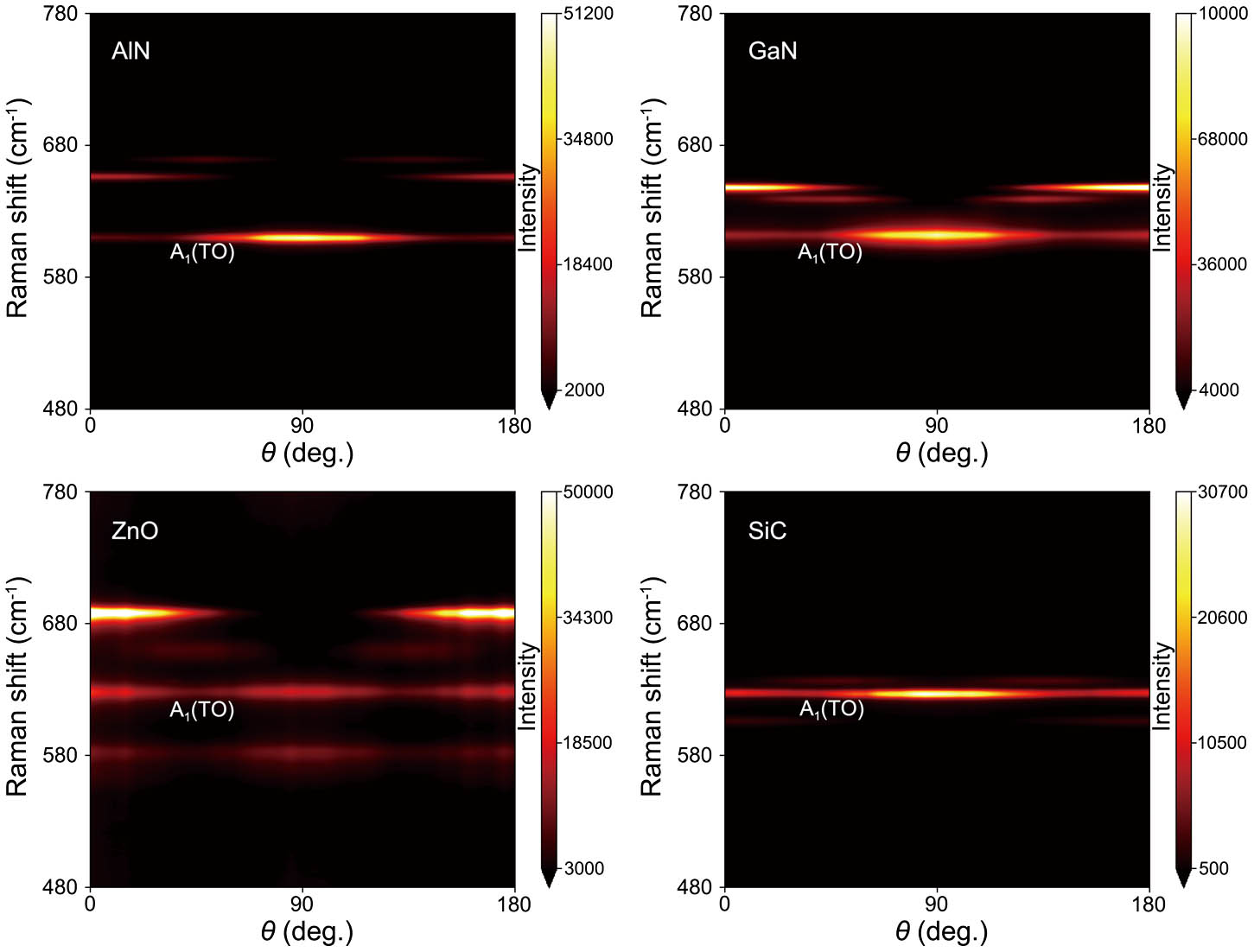
The so-called “phase difference” is commonly introduced as a phenomenological parameter in Raman tensor theory, so as to fit the experimental data well. Although phase difference is widely recognized as an intrinsic property of crystals, its physics still remains ambiguous. Recently, Kranert et al. have presented a new formalism to explain the origin of phase difference theoretically. Here, we systematically conducted experimental research with polar phonons in wurtzite crystals, the results of which strongly suggest that the phase difference should be predetermined in a Raman tensor, rather than be treated as Raman tensor elements traditionally or as an intrinsic property. On the grounds of pinpointing existing logical flaws in Raman tensor study, we provide a logically clear paradigm.
Scattering, Raman Spectroscopy, Raman Photonics Research
2018, 6(7): 07000709

Author Affiliations
Abstract
1 State Key Laboratory of Transient Optics and Photonics, Xi’an Institute of Optics and Precision Mechanics, Chinese Academy of Science (CAS), Xi’an 710119, China
2 University of Chinese Academy of Sciences (CAS), Beijing 100049, China
3 State Key Laboratory of Power Systems, Department of Thermal Engineering, Tsinghua-BP Clean Energy Center, Tsinghua University, Beijing 100084, China
4 School of Science, Xi’an Jiaotong University, Xi’an 710049, China
In this Letter, a miniature wearable Raman spectroscopy system is developed. A wearable fiber-optic probe is employed to help the stable and convenient collection of Raman spectra. A nonlinear partial least squares model based on a multivariate dominant factor is employed to predict the glucose level. The mean coefficients of determination are 0.99, 0.893, and 0.844 for the glucose solution, laboratory rats, and human volunteers. The results demonstrate that a miniature wearable Raman spectroscopy system is feasible to achieve the noninvasive detection of human blood glucose and has important clinical application value in disease diagnosis.
300.6450 Spectroscopy, Raman 300.6190 Spectrometers 120.4290 Nondestructive testing Chinese Optics Letters
2017, 15(8): 083001
Author Affiliations
Abstract
1 School of Materials and Metallurgy, Northeastern University, Shenyang 110819, China
2 Engineering Research Center for Process Technology of Nonferrous Metallurgy, Ministry of Education, Shenyang 110819, China
We study ionic structure of KNO3–NaNO2 melts under air atmosphere by using Raman spectroscopy. Molar fraction of NO3- and NO2- is obtained and thermal stability of this kind of melts system is then analyzed. The results show that when the temperature is increased to a certain value, equilibrium between the decom-position of NO3- and the oxidation of NO2- exists in KNO3–NaNO2 melts. When temperature is higher than 644 K, the molar fraction of NO3- decreases a little with temperature increasing for the melts in which the initial fraction of KNO3 is 90 wt%, but for the melts in which the initial fraction of KNO3 is 10–80 wt%, the molar fraction of NO3- increases with temperature, and the increasing rate is slower for a higher initial frac-tion of KNO3. Molar fraction of NO3- increment increases linearly with initial fraction of NaNO2. The sample in which the initial fractions of NaNO2 are 11.3 and 14.5 wt% under air atmosphere shows the best thermal stability at 762 and 880 K, respectively.
300.6450 Spectroscopy, Raman 290.5860 Scattering, Raman Chinese Optics Letters
2014, 12(9): 093001
Author Affiliations
Abstract
We develop optical fiber nanoprobe by spark fused taper and acid corrosion methods. By coupling with 3-aminopropyltrimethoxysilane, gold nanoparticles are solidified onto the surface of fiber optic and then the optical fiber sensor is prepared using surface-enhanced Raman spectroscopy (SERS) measurement of the cell solution. The SERS of the esophagus cancer cell solution is then measured by direct detection and fiber detection methods, and the relationship between SERS fiber detection and the length of optical fiber ensor is studied. This is helpful for the SERS measurement of tissues and organs using the optical fiber sensor.
300.6450 Spectroscopy, Raman 300.6390 Spectroscopy, molecular 300.6360 Spectroscopy, laser Chinese Optics Letters
2014, 12(s1): S13001
Author Affiliations
Abstract
A porous silicon microcavity (PSM) is highly sensitive for sensing applications due to its high surface area and a narrow resonance peak. In this letter, we fabricated the PSM by alternate current density from a low value to a high value during double-tank electrochemical anodization at different electrolyte temperatures. Results show that with the increase of the electrolyte temperature, the rate of the PS etching becomes faster and the refractive index of the PS layer becomes smaller. The thickness of the PS increases faster than the decrease of the refractive index of the PS.
240.6695 Surface-enhanced Raman scattering 160.4670 Optical materials 160.3900 Metals 300.6450 Spectroscopy, Raman Chinese Optics Letters
2014, 12(s1): S12402
Author Affiliations
Abstract
A simple, low-cost, and high-efficient method is used for the fabrication of surface-enhanced Raman scattering (SERS) substrates. Silver particles deposited on porous silicon are prepared as a highly efficient SERS substrate by direct immersion of porous silicon in silver solution. The SERS measured with rhodamine 6G as a target molecule is affected by the morphology of silver particles on the top of porous silicon layer. The effect of solution concentration, dipping time, and thickness of porous layer on the morphology of silver particle is investigated. Highly efficient SERS spectra are observed for substrates with porous layer thickness of about 3 μm and incubated in the 50 mM AgNO3 solution for 3 minutes. The SEM images of the substrates show that there are many small Ag particles with the size of a few nanometers among large Ag particles with the size of several microns.
240.6695 Surface-enhanced Raman scattering 160.4670 Optical materials 160.3900 Metals 300.6450 Spectroscopy, Raman Chinese Optics Letters
2014, 12(s1): S12401
Author Affiliations
Abstract
Cephalosporins are widely used as veterinary and human antibiotics. However, cephalosporin abuse is harmful to human health and causes allergic reactions or antibiotic resistance. We investigate a method featuring Raman spectroscopy and chemometrics to quantify mixture solutions of three typical cephalosporins, namely, ceftriaxone, cefotaxime sodium, and cefazolin sodium. Partial least-squares regression models are built on spectral data that are preprocessed by various methods. With prediction relative errors within 5% and high correlation coefficients of 0.998, we demonstrate that Raman spectroscopy combined with multivariate analysis is feasible for use in the quantitative determination of cephalosporin solutions.
300.6450 Spectroscopy, Raman 070.4340 Nonlinear optical signal processing 070.4790 Spectrum analysis Chinese Optics Letters
2013, 11(12): 123001
Author Affiliations
Abstract
Graphene oxide (GO)/Ag nanoparticle (NP) hybrids are obtained by in situ reduction of Ag NPs on GO sheets. In this letter, the influence of the conformation of GO sheets on the surface-enhanced Raman scattering (SERS) effect of GO/Ag NPs is investigated by covalently grafting folic acid (FA) molecules onto graphite sheets. SERS measurements are conducted in aqueous solutions with different pH values. Data show that the SERS signals of FA are pH dependent, consistent with the morphological changes of GO sheets.
300.6450 Spectroscopy, Raman 160.4236 Nanomaterials Chinese Optics Letters
2013, 11(8): 083001


